https://t.co/ZLzH00ba8Q
“Men . . . go mad in herds, while they only recover their senses slowly, and one by one.”
Mijn dagelijkse portie 'sanity' and 'mental health'
#coronamaatregelen #coronavirus #vaccination
"Except. For. One. That’s total elimination of the disease."
"That may be close to impossible and incredibly expensive to even try, but like eliminating all carbon emissions in a decade it is a goal."
Social distancing dividers at various businesses and schools are just like the sneeze guards at the local buffet."
And now it’s being threatened by a shift of resources to, you guessed it, Covid-19."
Imagine throwing a brick into an Olympic-sized pool and trying to measure a rise in the waterline."
Perhaps, but obviously some lives matter more than most."
Ik had het niet beter kunnen omschrijven.
More from Health


Sarcomeres in cardiac myocytes (heart muscle cells) are mechanically coupled to focal adhesions through dorsal stress fiber-like structures. #cardiotwitter #CellBiology
1/13
A thread based on Figure 1
A mature adult cardiac myocyte is packed with sarcomeres, whose contractile forces are coupled to the extracellular environment. With sarcomeres so close to the plasma membrane, how can we study the nature of this coupling?
2/13
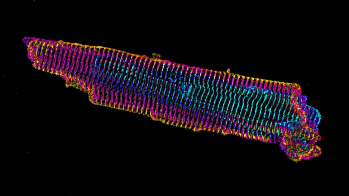
Short answer: find a model system where the sarcomeres are not so close to what the cardiac myocyte is attached to. Enter, iPS cell-derived cardiac myocytes. These are “immature” in culture as they resemble fetal or neonatal cardiac myocytes.
3/13
Our previous work on iPS cardiac myocytes reported that sarcomere containing myofibrils assembled on the top surface of the myocyte.
https://t.co/xIBCu3hG1W
4/13
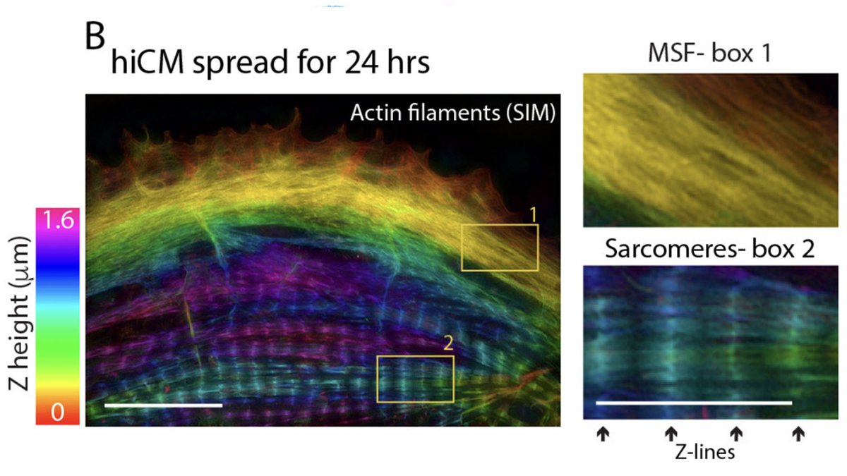
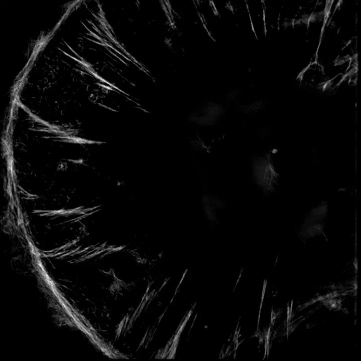
The sarcomeres seemed to be connected to focal adhesions on the bottom of the cell by thin actin bundles that resembled the dorsal stress fibers (DSF) commonly found in non-muscle cells. This movie steps through a Z stack of a myocyte starting at the bottom of the cell.
5/13
1/13
A thread based on Figure 1
A mature adult cardiac myocyte is packed with sarcomeres, whose contractile forces are coupled to the extracellular environment. With sarcomeres so close to the plasma membrane, how can we study the nature of this coupling?
2/13
Short answer: find a model system where the sarcomeres are not so close to what the cardiac myocyte is attached to. Enter, iPS cell-derived cardiac myocytes. These are “immature” in culture as they resemble fetal or neonatal cardiac myocytes.
3/13
Our previous work on iPS cardiac myocytes reported that sarcomere containing myofibrils assembled on the top surface of the myocyte.
https://t.co/xIBCu3hG1W
4/13
The sarcomeres seemed to be connected to focal adhesions on the bottom of the cell by thin actin bundles that resembled the dorsal stress fibers (DSF) commonly found in non-muscle cells. This movie steps through a Z stack of a myocyte starting at the bottom of the cell.
5/13