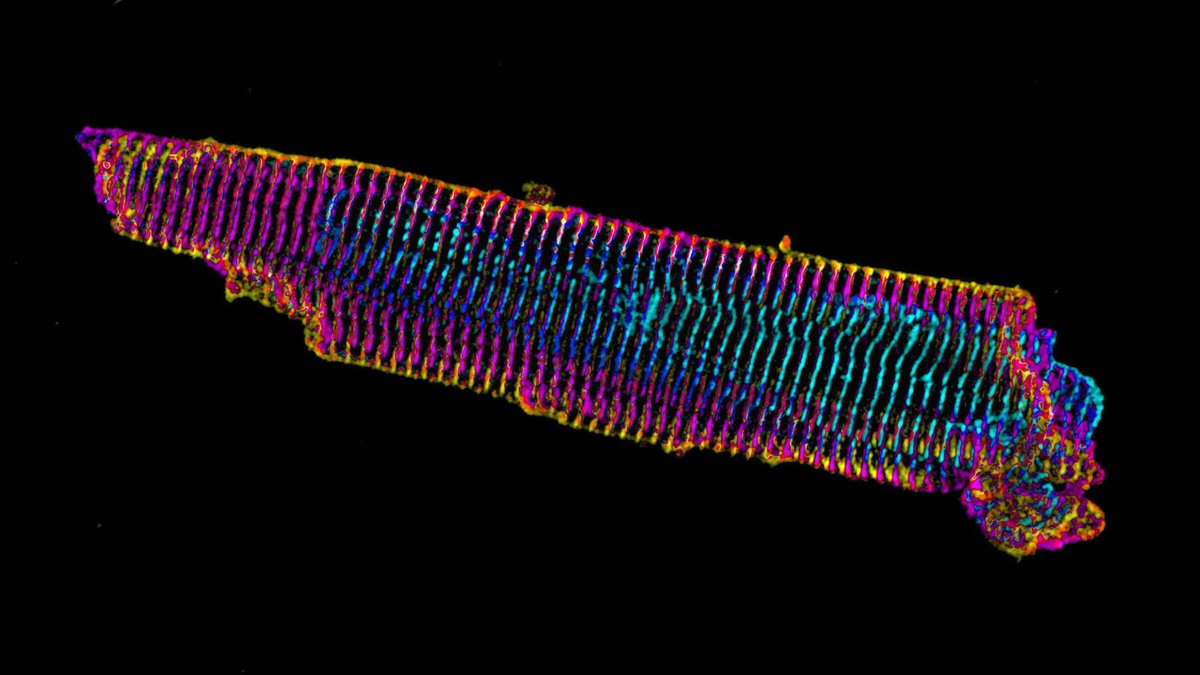
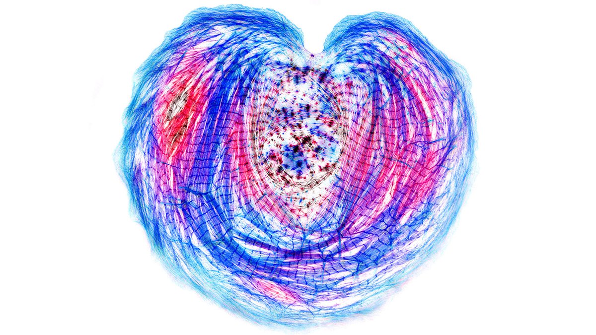
Sarcomeres in cardiac myocytes (heart muscle cells) are mechanically coupled to focal adhesions through dorsal stress fiber-like structures. #cardiotwitter #CellBiology
1/13
A thread based on Figure 1

A mature adult cardiac myocyte is packed with sarcomeres, whose contractile forces are coupled to the extracellular environment. With sarcomeres so close to the plasma membrane, how can we study the nature of this coupling?
2/13
Short answer: find a model system where the sarcomeres are not so close to what the cardiac myocyte is attached to. Enter, iPS cell-derived cardiac myocytes. These are “immature” in culture as they resemble fetal or neonatal cardiac myocytes.
3/13
Our previous work on iPS cardiac myocytes reported that sarcomere containing myofibrils assembled on the top surface of the myocyte.
https://t.co/xIBCu3hG1W
4/13
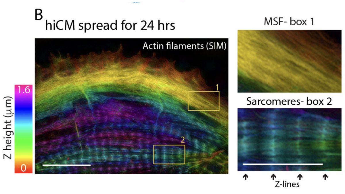
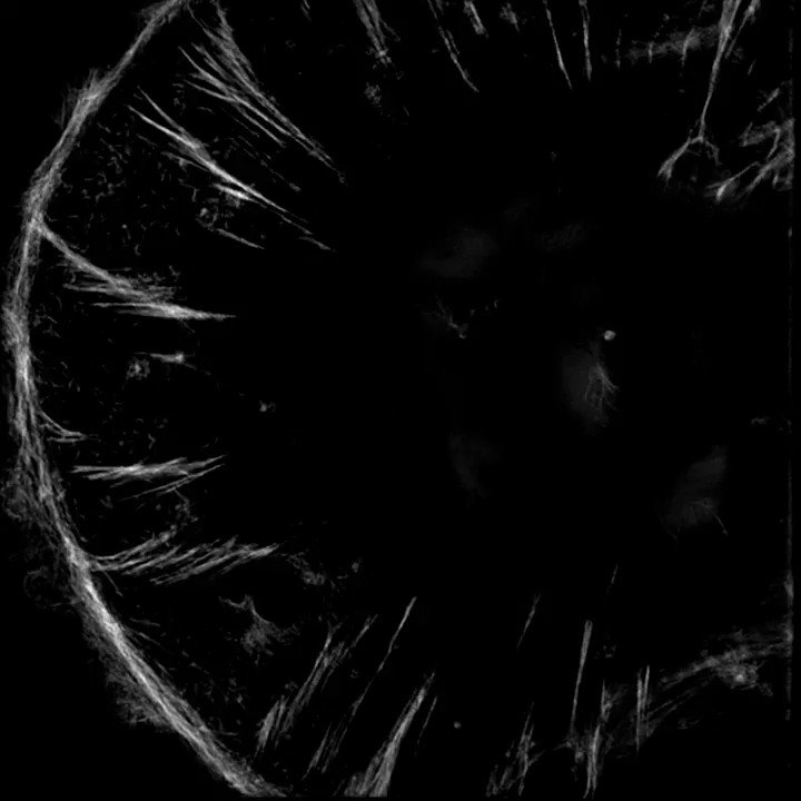
The sarcomeres seemed to be connected to focal adhesions on the bottom of the cell by thin actin bundles that resembled the dorsal stress fibers (DSF) commonly found in non-muscle cells. This movie steps through a Z stack of a myocyte starting at the bottom of the cell.
5/13
Another way of visualizing dorsal stress fibers. This montage steps through the edge of a myocyte in the Z dimension. Arrows denote individual dorsal stress fibers.
6/13
Another way of visualizing the paths of the dorsal stress fibers is by using a color overlay.
7/13
In non-muscle cells, dorsal stress fibers can mechanically couple adhesions to the substrate to stress fibers on top of the cell.
This led us to this hypothesis: dorsal stress fibers also mechanically couple focal adhesions and sarcomeres. But how to test it? Answer- LASERS!
8/13
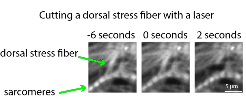
If the contractile forces generated by the sarcomeres are balanced by attachment to focal adhesions, then we reasoned that cutting a dorsal stress fiber would result in the sarcomeres rabidly moving (i.e., snapping) away from the adhesions.
Enter: laser ablation.
9/13
Cutting a dorsal stress fiber did make the sarcomeres it was attached to move away from the edge of the cardiac myocyte. What? Don’t you see that it moved? Maybe we should look at it in a different—animated—way.
10/13
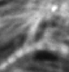
This gif shows just before (0s) and just after (2s) the dorsal stress fiber was cut. The movement of the sarcomeres was clear but subtle, so we sought to test the mechanical connection in another way.
Enter: cutting sarcomeres to test if dorsal stress fibers move.
11/13
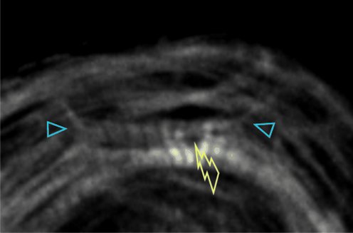
If the sarcomeres and dorsal stress fibers are mechanically coupled, then cutting the sarcomeres should result in the movement of the dorsal stress fibers. It did.
Blue arrowheads show the dorsal stress fibers.
12/13
So what is the implication of the mechanical coupling between sarcomeres and dorsal stress fibers?
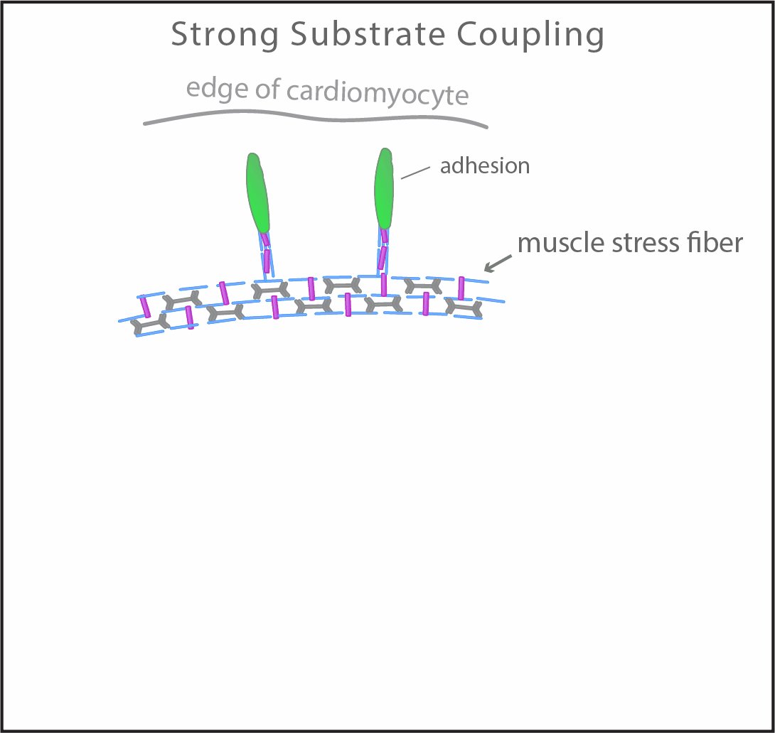
Our interpretation from 8 additional data figures: Strong coupling facilitates the maturation of sarcomeres. Weak coupling slows maturation down.
13/13
https://t.co/cKnT7wXo0w

More from Health

Thread on how atheism leads to mental retardation (backed with medical citations🧵💉)

To start with, atheism is an unnatural self-contradicting doctrine.

Medical terminology proves that human beings are naturally pre-disposed to believe in God. Oxford scientists assert that people are "born believers".

https://t.co/kE0Fi588yn
https://t.co/OqyXcGIMJn


It should be known that atheism could never produce an intelligently-functioning society and neither ever will.

Contrastingly, Islam produced several intellectuals & polymaths, was on the forefront of scientific development, boasting 100% literacy


It is also scientifically proven that atheism led to lesser scientific curiosity and scientific frauds, which is also why atheists incline to pseudo-science.

Whereas, religion in general and Islam in particular boosted education.

https://t.co/19Onc84u3g


Atheists are also likely to affected by pervasive mental and developmental disorders like high-functioning autism.

Cognitive Scientists and renowned Neurologists found that more atheism is leads to greater autism.

https://t.co/zRjEyFoX3P

You May Also Like