ADHD, a thread (1)
If you have #ADHD & are struggling please know you’re not alone. This week on @Overview, we're talking about living w/ ADHD & Anxiety during the pandemic & many the stigmas & misconceptions that making things worse.
https://t.co/RO1ZGTlMq6


https://t.co/TXL9aOebDj
Like, right now, I’m furiously locked into this thread, & breaking away is super disconcerting & almost painful. When honed & controlled it can supercharge your productivity. But when unchecked it can feel like this.(12)

Quick warning, my choices and perspective don’t reflect a “right” or “wrong” way to do things. I believe everyone should do what works for best for them. (14)
https://t.co/PInu3lt8mi
https://t.co/1jJe4K02F9
https://t.co/7L6NJjSbA9
1. I want to talk about ADHD. I was diagnosed with ADD when I was 13-years-old. To say it's misunderstood (both by people who have ADHD and people who don't have it) is an understatement. What people need to understand about those of us who have ADHD is we are not homogeneous
— Yashar Ali \U0001f418 (@yashar) February 26, 2019
https://t.co/1jJe4K02F9
More from Health




Sarcomeres in cardiac myocytes (heart muscle cells) are mechanically coupled to focal adhesions through dorsal stress fiber-like structures. #cardiotwitter #CellBiology
1/13
A thread based on Figure 1
A mature adult cardiac myocyte is packed with sarcomeres, whose contractile forces are coupled to the extracellular environment. With sarcomeres so close to the plasma membrane, how can we study the nature of this coupling?
2/13
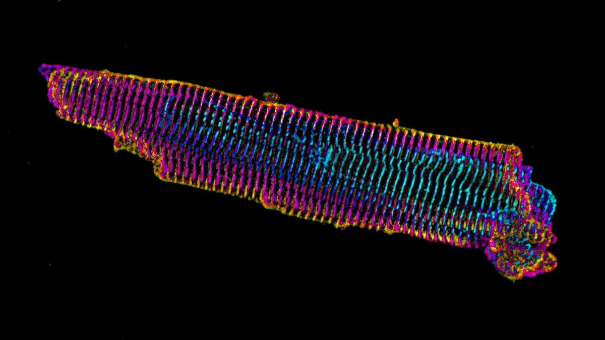
Short answer: find a model system where the sarcomeres are not so close to what the cardiac myocyte is attached to. Enter, iPS cell-derived cardiac myocytes. These are “immature” in culture as they resemble fetal or neonatal cardiac myocytes.
3/13
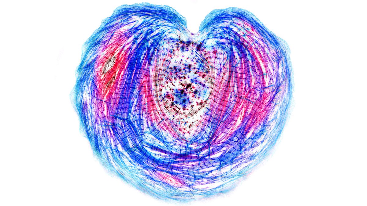
Our previous work on iPS cardiac myocytes reported that sarcomere containing myofibrils assembled on the top surface of the myocyte.
https://t.co/xIBCu3hG1W
4/13
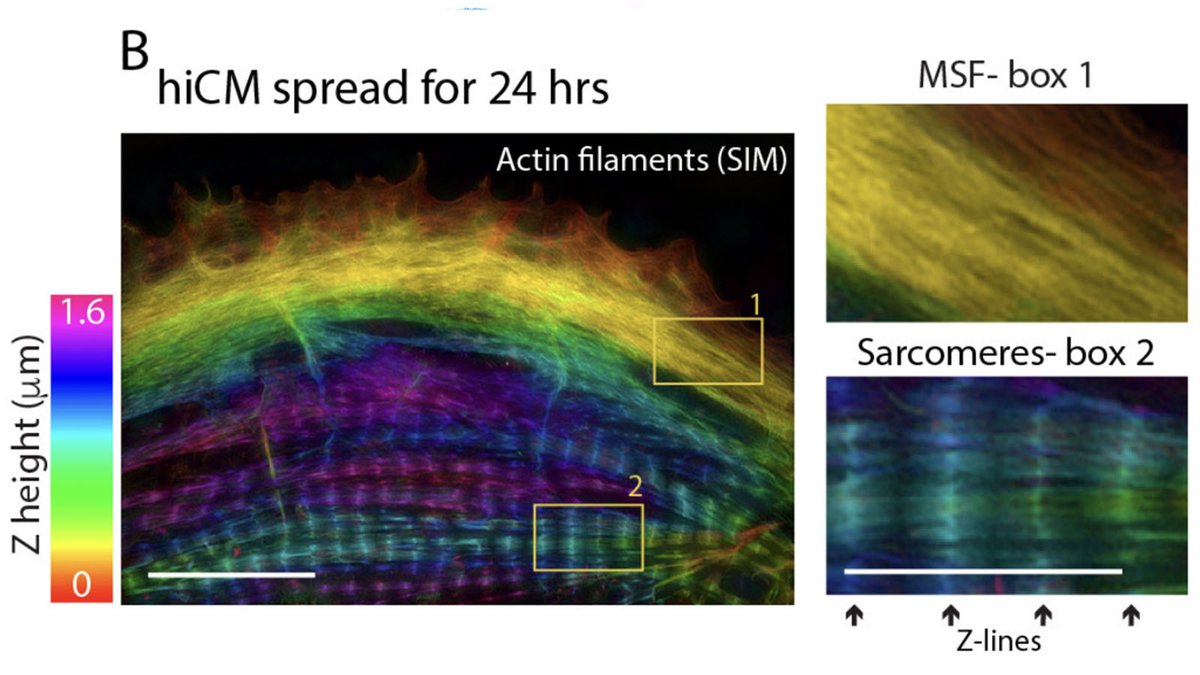
The sarcomeres seemed to be connected to focal adhesions on the bottom of the cell by thin actin bundles that resembled the dorsal stress fibers (DSF) commonly found in non-muscle cells. This movie steps through a Z stack of a myocyte starting at the bottom of the cell.
5/13
1/13
A thread based on Figure 1
A mature adult cardiac myocyte is packed with sarcomeres, whose contractile forces are coupled to the extracellular environment. With sarcomeres so close to the plasma membrane, how can we study the nature of this coupling?
2/13
Short answer: find a model system where the sarcomeres are not so close to what the cardiac myocyte is attached to. Enter, iPS cell-derived cardiac myocytes. These are “immature” in culture as they resemble fetal or neonatal cardiac myocytes.
3/13
Our previous work on iPS cardiac myocytes reported that sarcomere containing myofibrils assembled on the top surface of the myocyte.
https://t.co/xIBCu3hG1W
4/13
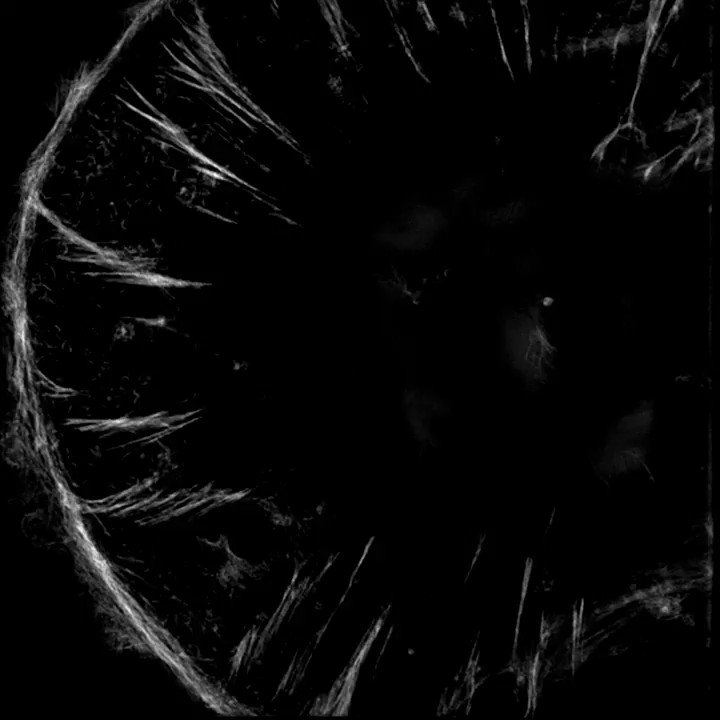
The sarcomeres seemed to be connected to focal adhesions on the bottom of the cell by thin actin bundles that resembled the dorsal stress fibers (DSF) commonly found in non-muscle cells. This movie steps through a Z stack of a myocyte starting at the bottom of the cell.
5/13